Balancing Energy and Cellular Recovery: The Clinical Reality of Advanced Dermal Remodeling
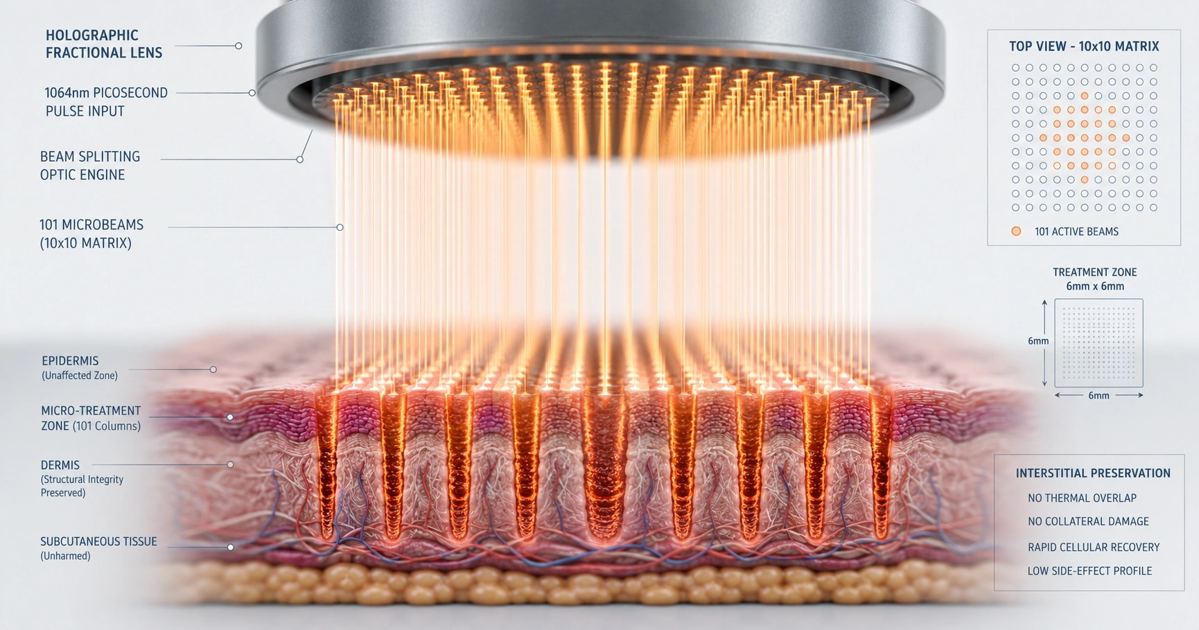
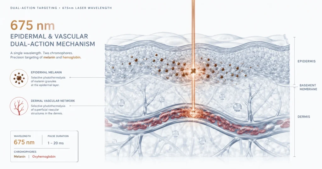
This article examines the clinical safety, technical parameters, and longitudinal outcomes of a 1064 nm picosecond-domain laser utilizing a fractionated holographic microlens array for facial melasma management. Clinical trial data demonstrates a highly significant mean modified melasma area and severity index (mMASI) reduction of 2.1 units, stabilized at both the 3-month and 8-month post-treatment milestones. The protocol leverages high peak power and low fluences to target melanin pigment while strictly limiting the post-treatment inflammatory triggers that typically cause secondary pigment recurrence.
High Peak Power Delivery and Laser Pigmentation Management
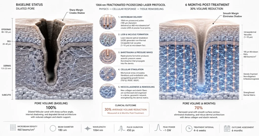
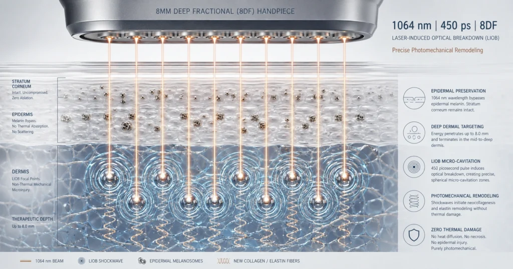
Modern clinical protocols manage facial hyperpigmentation by delivering high peak powers with relatively low fluences to target melanin pigment while minimizing post-treatment inflammation. Traditional laser modalities rely heavily on extended thermal accumulation, which introduces a known risk of reactive melanocyte activation and subsequent pigment rebound. Picosecond-domain laser systems alter this target interaction by utilizing ultra-short 450 picosecond pulses.
This rapid delivery speed targets melanin pigment effectively while minimizing subsequent inflammation. Restricting localized heat accumulation is essential because melasma functions primarily as an inflammatory-modulated condition that is easily exacerbated by thermal stress. Furthermore, this fractionated 1064 nm approach avoids the specific risks of long-lasting or permanent hypopigmentation frequently documented with traditional low-fluence, open-beam nanosecond laser toning treatments.
The Holographic Matrix and Interstitial Tissue Preservation
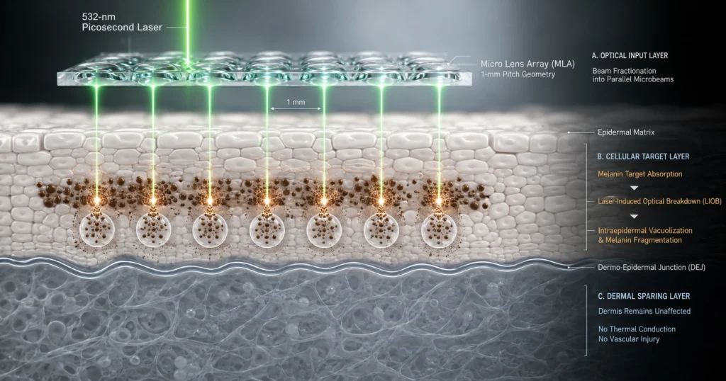
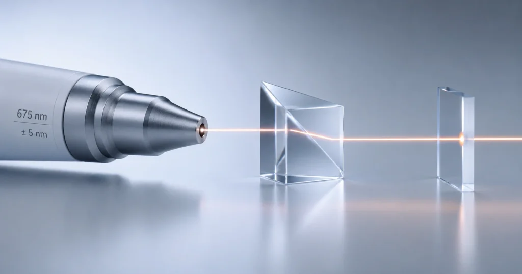
A fractionated holographic microlens array splits laser energy into a precise microbeam array to limit total skin exposure and protect surrounding tissue architecture. The holographic manufacturing process creates a specialized beam-splitting handpiece that delivers a 10×10 fractional matrix composed of 101 distinct microbeams, each measuring 150 µm in diameter, arranged within a 6×6 mm square treatment area.
Testing with highly sensitive laser burn paper confirms that there is zero energy effect in the interstitial tissue zones between these 101 sharp, well-demarcated microbeams. This complete spatial isolation confines high-intensity energy to micro-treatment zones. Leaving the adjacent skin completely unexposed limits total localized trauma, resulting in low side-effect profiles and supporting orderly dermal remodeling.
Sub-Surface Dermal Inflammatory Responses and Longitudinal Efficacy
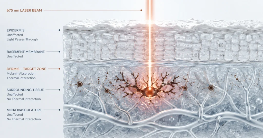
Deper laser penetration paths trigger a sub-surface dermal inflammatory response that addresses the complex, multi-layered pathology of facial hyperpigmentation. Melasma pathology extends beyond superficial melanocytes to involve interactions between keratinocytes, cutaneous nerves, dermal mast cells, neovascularization, and inflammatory mediators. Superficial deposition of fractionated laser energy generates a localized dermal inflammatory response, clinically evident as transient erythema.
Because inflammatory cells originate within the dermal vasculature, this targeted, sub-lethal cascade prompts structural cellular remodeling that drives pigment clearance across epidermal, dermal, and mixed melasma variants. Clinical trial tracking of 20 subjects with Fitzpatrick skin types III–VI demonstrated substantial, documented outcomes:
- Independent, blinded dermatologists correctly identified true post-treatment images in 80% of randomized patient cases.
- Blinded visual evaluations demonstrated a 37% mean objective improvement at 3 months and a 27% mean improvement at 8 months post-treatment.
- The average mMASI score showed a highly significant reduction from a baseline mean of 5.90 ± 2.86 down to a stabilized mean of 3.78 ± 3.05 at the final 8-month follow-up milestone (p = 0.004).
- Measured mMASI score improvements were successfully achieved in 72% of subjects at 3 months and 71% of subjects at 8 months.
Multi-Modal Integration and Cellular Recovery Protocols
Comprehensive melasma clearance profiles rely on nesting fractionated picosecond laser treatments as clinical adjuncts within a daily multi-modal management regimen. Melasma is a chronic, acquired disorder characterized by frequent exacerbations, making its long-term management challenging due to high natural recurrence rates. Longitudinal tracking indicates a natural recurrence trajectory over extended follow-up windows across all light and laser modalities, proving that lasers manage rather than permanently cure the condition.
To stabilize and preserve clinical clearance rates, the laser protocol must be integrated with a structured daily home routine:
- Topical Agents: Daily application of topical retinoids, hydroquinone, azelaic acid, kojic acid, corticosteroids, or topical tranexamic acid to regulate pigment pathways.
- Photoprotection Architecture: Continuous use of broad-spectrum sunscreens incorporating physical and chemical blockers to protect against ultraviolet B (UVB), ultraviolet A (UVA), and long-wavelength visible light.
- Environmental Barrier Shielding: Installation of clear, ultraviolet-absorbing car window film to eliminate the 70% of UVA radiation that readily penetrates standard vehicle glass during daily transit.
Quantifying Post-Treatment Sequelae and Patient Tolerability
The fractionated 1064 nm picosecond-domain laser protocol exhibits a favorable safety profile with mild, predictable, and entirely self-limiting post-treatment responses. The procedure is well tolerated, recording a mean discomfort score of 3.8 ± 2.3 on an 11-point numerical pain rating scale. This high tolerability profile allows clinical application without topical anesthetic agents.
Immediate post-procedure sequelae remain strictly temporary:
- Erythema: Present in 100% of treatments immediately post-procedure, categorized as mild in 61.2% of cases and moderate in 38.8%.
- Edema: Observed in 40% of treatments, presenting exclusively as mild swelling.
- Petechiae: Occurs in a low 2.5% of total treatments and resolves fully within 1 to 2 days.
- Long-Term Safety: Tracking confirms zero instances of permanent scarring, post-inflammatory hyperpigmentation, or mottled hypopigmentation across the longitudinal evaluation period.
References
Bernstein EF, Basilavecchio LD, Wang J. Melasma treatment with a 1064 nm, picosecond-domain laser with a fractionated multibeam lens array. Lasers in Surgery and Medicine. 2023;55(9):801-808. doi:10.1002/lsm.23723.