The Molecular Architecture of Skin Tightening: How 6.78 MHz Monopolar Radiofrequency Modulates Collagen Gene Expression
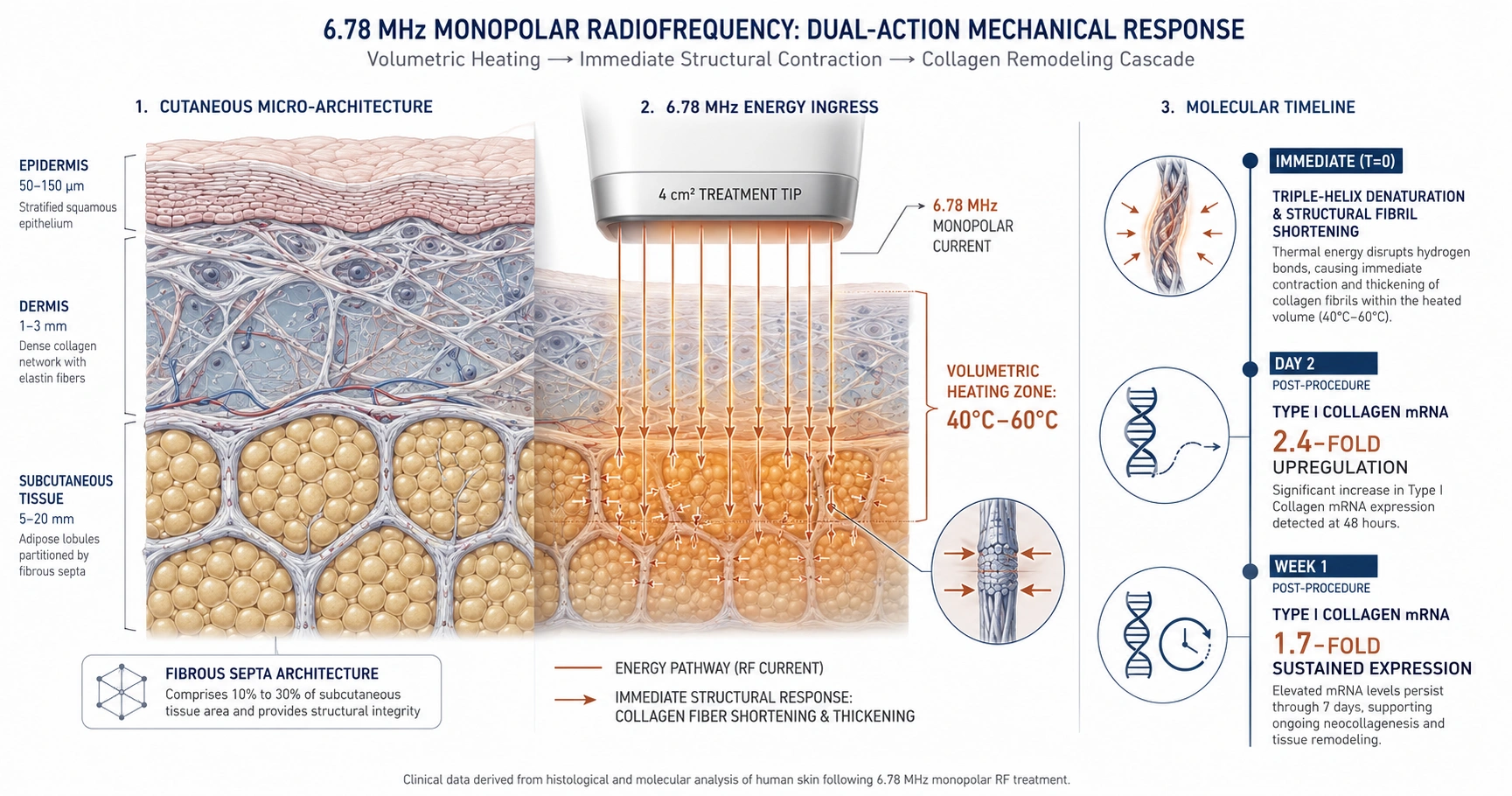
Clinical data indicates that 6.78 MHz monopolar radiofrequency addresses lower facial laxity via a two-phase physiological mechanism. Immediate volumetric heating of deep tissue layers induces collagen fibril contraction. This thermal stimulus triggers a natural wound-healing cascade, resulting in a documented 2.4-fold upregulation of Type I collagen mRNA steady-state expression by post-procedure Day 2. Blinded evaluations confirm that 86.67% of subjects maintain objective clinical improvement at six months.
Biophysics of 6.78 MHz Monopolar Radiofrequency
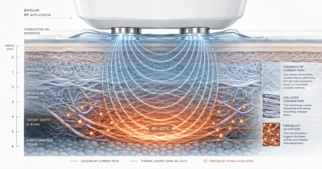
The 6.78 MHz monopolar radiofrequency system delivers high-frequency electrical current via a 4 cm² transducer tip through targeted anatomical zones to a grounding pad. This closed circuit generates deep-tissue volumetric heating between 40°C and 60°C within the dermis and subcutaneous architectures. Energy systematically flows through collagen-based fibrous septa, which constitute 10% to 30% of subcutaneous tissue. This localized thermal energy induces immediate triple-helix collagen denaturation, resulting in fibril shortening, tissue thickening, and immediate visible contouring.
Genetic Modulation and Neocollagenesis Timeline
Beyond immediate physical contraction, the thermal profile initiates a long-term tissue remodeling cascade. The controlled heating stimulates fibroblasts, altering collagen gene expression without inducing permanent structural injury. Clinical analysis confirms a significant shift in molecular architecture post-procedure:
- Day 2 Post-Procedure: Steady-state expression of Type I collagen mRNA increases 2.4-fold compared to baseline levels.
- Week 1 Post-Procedure: A 1.7-fold higher steady-state expression of Type I collagen mRNA is sustained within the treated tissue.
This genetic upregulation drives neocollagenesis, rebuilding the dermal matrix progressively over subsequent months.
Longitudinal Efficacy and Safety Profile
In a prospective clinical cohort study monitoring Asian subjects with mild-to-moderate lower facial laxity, objective clinical outcomes were tracked using a 6-point improvement scale. Blinded, independent board-certified dermatologists recorded structural improvements at key temporal intervals:
- 3-Month Checkpoint: 40.00% of subjects demonstrated moderate and 3.33% demonstrated marked tissue improvement.
- 6-Month Checkpoint: 86.67% of subjects maintained objective clinical improvement, demonstrating statistical progression from earlier follow-ups (P < 0.01).
Epidermal safety is maintained via integrated real-time contact pressure sensors, continuous skin impedance monitoring, and a multi-phase cryogen gas cooling system that shields surface tissue before, during, and after each energy pulse. The procedure yielded a mean pain score of 3.13 out of 10. Transient post-treatment erythema occurred in 83.3% of subjects and resolved naturally within 1 to 2 days, allowing an immediate return to regular activities.
References
Wanitphakdeedecha R, Yogya Y, Yan C, Phumariyapong P, Nanchaipruek Y, Thongjaroensirikul P, Techapichetvanich T, Eimpunth S, Manuskiatti W. Efficacy and Safety of Monopolar Radiofrequency for Treatment of Lower Facial Laxity in Asians. Dermatol Ther (Heidelb). 2022;12:2563-2573. https://doi.org/10.1007/s13555-022-00817-8