The Cellular Science of Epidermal Melanin Regulation: Understanding Laser-Induced Optical Breakdown (LIOB)
This article examines the clinical application of a 532-nm fractionated picosecond laser utilizing a high-coverage micro lens array for managing localized epidermal dyspigmentation. By inducing laser-induced optical breakdown (LIOB), this methodology targets epidermal melanin while restricting thermal energy transfer to the deeper dermis. Quantitative data demonstrates documented pigment clearance at 12 weeks post-treatment alongside low patient discomfort scores and rapid recovery timelines.
Characterizing Epidermal Dyspigmentation: Facial PUL
Facial partial unilateral lentiginosis (PUL) is an skin pigmentation disorder characterized by multiple clustered brown macules with a distinct demarcation at the midline. Pre-treatment diagnostics utilize polarized and non-polarized dermoscopy to identify the structural presence of an accentuated, regular pseudo-reticular pigment network. This precise evaluation ensures the target chromophore is accurately mapped prior to initiating specific laser energy application protocols.
The Physics of 532-nm Fractionated Picosecond Delivery
Wavelength Selectivity and Micro Lens Array Geometry
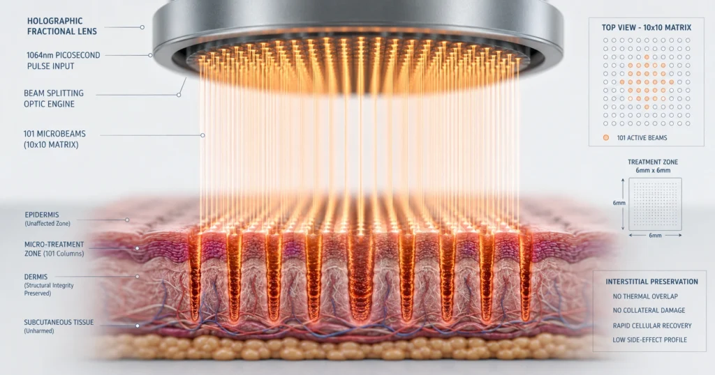
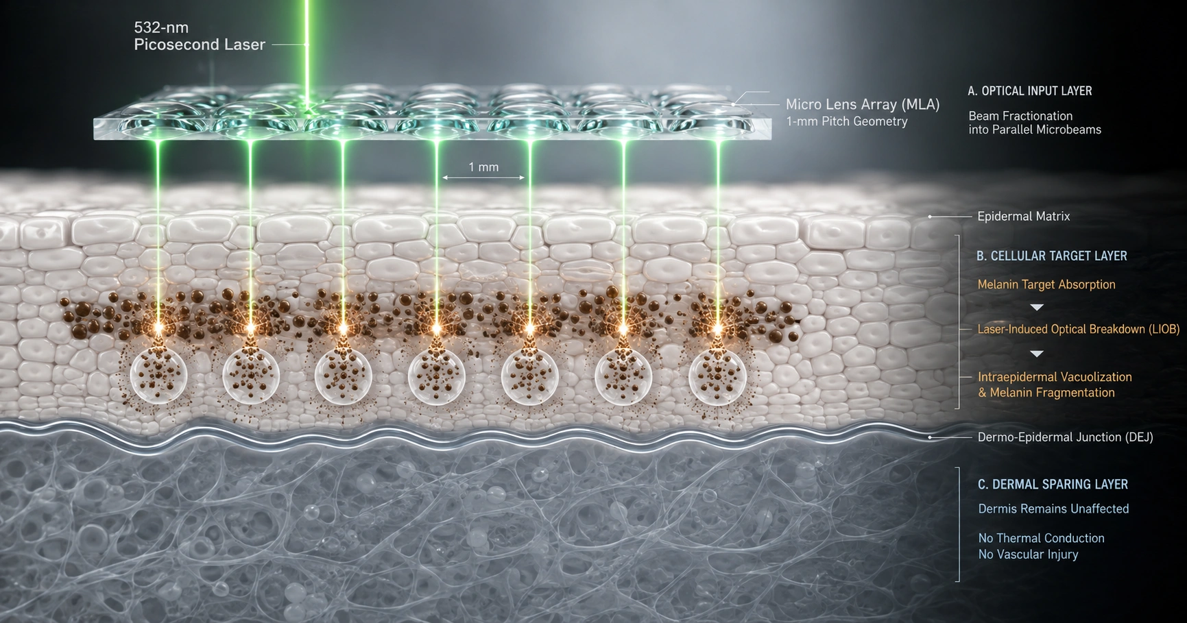
Targeting superficial dyspigmentation requires the selection of a 532-nm Nd:YAG wavelength due to its high absorption coefficient for melanin. To control energy delivery, a specialized 9 mm high-coverage micro lens array (MLA) handpiece fractionates the solid laser beam into individual microbeams. The resulting microdots measure 0.5 mm in diameter with a 1 mm inter-dot distance, covering exactly 20% of the active treatment area to preserve surrounding tissue.
The Physiological Mechanism of LIOB
Intraepidermal Vacuolization and Plasma Confinement
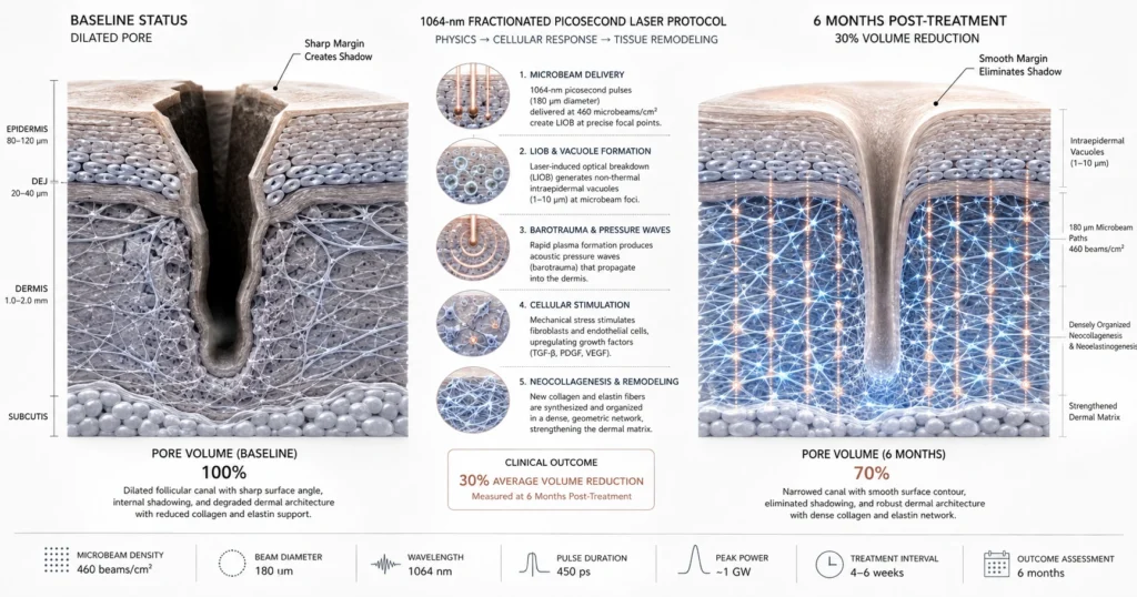
Laser-induced optical breakdown (LIOB) serves as the primary mechanism for fragmented pigment resolution. Ultra-short picosecond pulses generate high-energy microbeams that induce focal intraepidermal vacuoles within the lower epidermis. Energy absorption occurs predominantly within the nearby plasma, which restricts thermal energy transfer into the dermis and mitigates the risk of post-inflammatory hyperpigmentation (PIH).
Standardized Clinical Protocol and Parameters
Reaching the designated clinical endpoint involves a structured protocol utilizing a fluence of 0.2–0.4 J/cm² delivered at a frequency of 2 Hz. The full treatment course consists of 4 sessions spaced at 4-week intervals, applying 2–3 passes with an approximate 10% overlap per pass. Pre-procedure care integrates a 45-minute topical anesthetic application, followed by immediate post-treatment ice cooling to regulate surface temperatures.
Quantifiable Clinical Outcomes and Safety Profile
The immediate clinical endpoint of this fractionated picosecond configuration is a honeycomb-pattern whitening of the pigment or mild erythema without petechiae. At a 12-week post-treatment follow-up, objective evaluations demonstrate significant clearance of the targeted pigmented macules. Patient tracking data confirms a satisfaction rating of 9 out of 10 and a minimal pain score of 2 out of 10. Expected side effects are strictly transient: mild erythema and burning sensations resolve within 1 hour post-treatment, and localized redness subsides completely within 3 to 5 days without scaling, purpura, or scarring.
References
Suparuj L, Therdpong T. Efficacy of 532-nm Nd: YAG Fractional Picosecond Laser with 9 mm High Coverage Handpiece Micro Lens Array in the Treatment of Facial Partial Unilateral Lentiginosis: A Case Report. J Lasers Med Sci. 2024;15:e24. doi:10.34172/jlms.2024.24.