The Scientific Metrics Behind a 30% Volumetric Pore Reduction
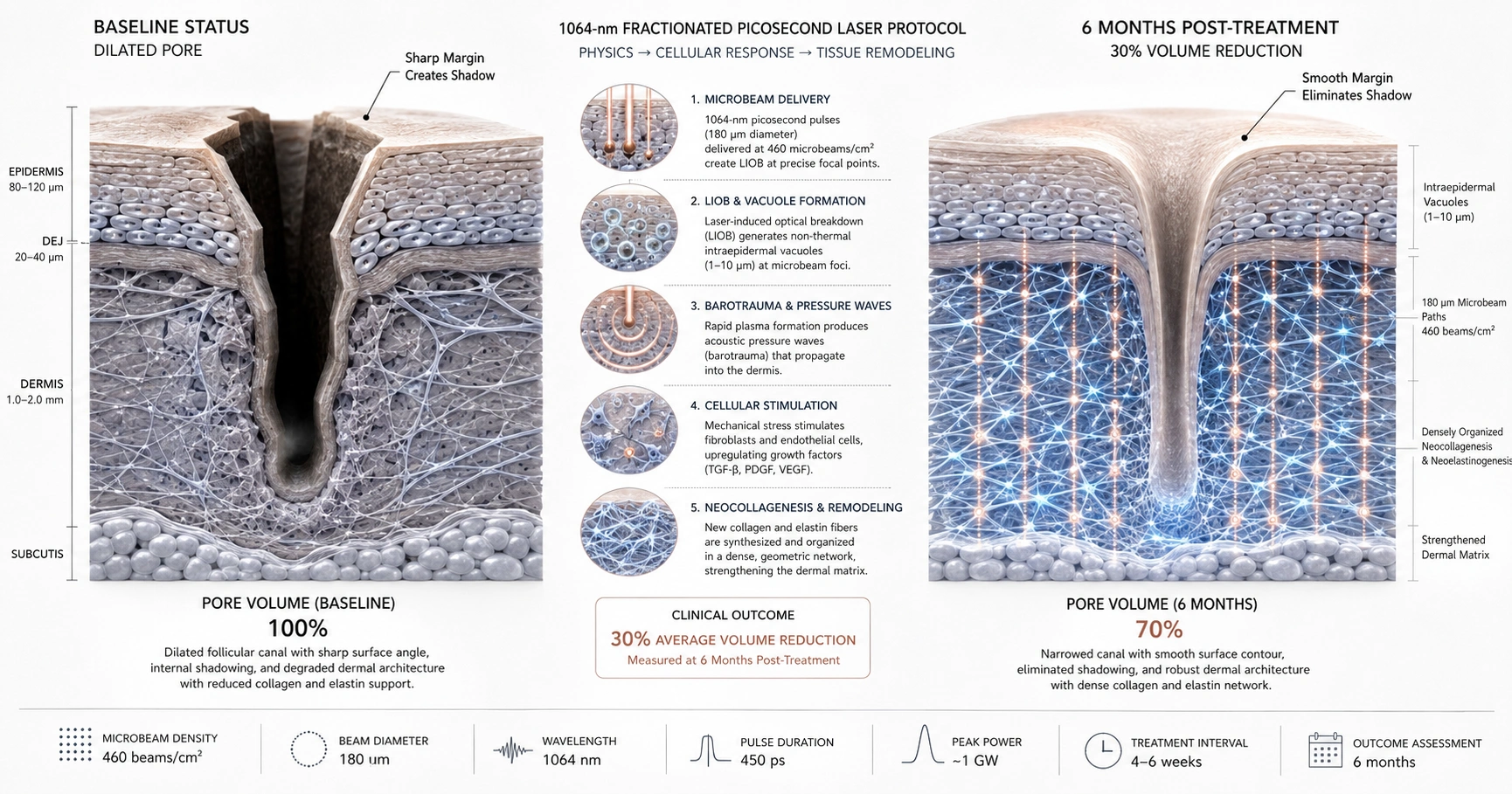
Clinical data from a prospective case-control study demonstrates that a three-session protocol using a 1064-nm picosecond laser with a fractionated microlens array achieves a statistically significant 30% average reduction in facial pore volume in Asian skin. Objective 3D topographic tracking confirms that mean pore metrics drop from a baseline of 1.15652 ± 0.614322 to 0.8087 ± 0.50515 at six months post-treatment. This structural refinement is mediated by non-thermal laser-induced optical breakdown (LIOB), which triggers targeted dermal remodeling and structural pore wall tightening.
Objective Measurement of Cutaneous Architecture
Evaluating pore refinement has historically relied on subjective clinical observation. To establish reproducible data, optical analysis using three-dimensional skin imaging technology mathematically records facial topography. In a clinical cohort of twenty-five patients with Fitzpatrick skin types III and IV, areas with visibly enlarged pores were mapped with translucent sheets to ensure structural consistency across longitudinal milestones. The resulting data sets revealed a significant reduction in total pore volume (p < 0.001) after three treatments spaced at four-week intervals.
The Physics of Laser-Induced Optical Breakdown
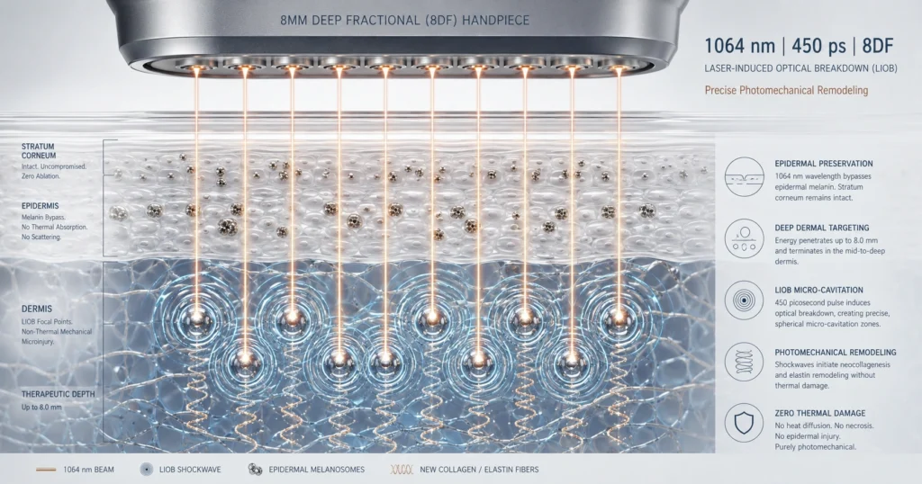
The structural tightening of the pilosebaceous follicle openings relies on targeted energy delivery within the ultra-short picosecond domain. Operating at a fluence of 0.8 J/cm² with an 8 mm spot size, a specialized microlens array (MLA) projects 460 microbeams per cm². This peak power is absorbed by intraepidermal melanin, causing an electron avalanche breakdown known as laser-induced optical breakdown (LIOB).
LIOB generates focal intraepidermal vacuoles in a non-thermal manner. The expansion and collapse of these localized zones produce acoustic pressure waves that propagate into the deeper tissue layers. This targeted dermal barotrauma modifies cellular signaling cascades, activating neocollagenesis and neoelastinogenesis. The progressive deposition of structured collagen fibers reinforces the slackened architectural walls of the pores, inducing clinical shrinkage and smoothing the perifollicular surface.
Chronological Trajectory and Safety Profiles
Unlike interventions causing temporary tissue edema, this mechanical remodeling undergoes an extended maturation process. Digital photography and blinded assessments by board-certified dermatologists show that topographical refinement continuously progresses between the 1-month and 6-month post-treatment milestones (p = 0.013). At the final six-month evaluation, 48% of subjects exhibited a moderate volume reduction (25% to 50%), and 28% demonstrated a marked reduction exceeding 50%.
The clinical protocol enforces parameter boundaries to manage cutaneous downtime in darker phototypes. Treatments are advanced until reaching a definitive tissue endpoint characterized by moderate erythema, mild-to-moderate edema, and mild purpura. Erythema and swelling persist for an average of 4 days, while micro-purpuric spots resolve within 5 to 7 days. Under standard topical anesthesia, the tolerability profile remains highly manageable, yielding a mean pain score of 3.205 ± 1.702 on a 1-to-10 scale. Crucially, zero instances of postinflammatory hyperpigmentation (PIH), hypopigmentation, or scar formation were documented. Minor transient acneiform eruptions occurred in 14% of sessions, resolving spontaneously within 24 to 48 hours.
References
Palawisuth S, Manuskiatti W, Apinuntham C, Wanitphakdeedecha R, Cembrano KAG. Quantitative assessment of the long-term efficacy and safety of a 1064-nm picosecond laser with fractionated microlens array in the treatment of enlarged pores in Asians: A case-control study. Lasers Surg Med. 2022;54:348-354. https://doi.org/10.1002/lsm.23449.