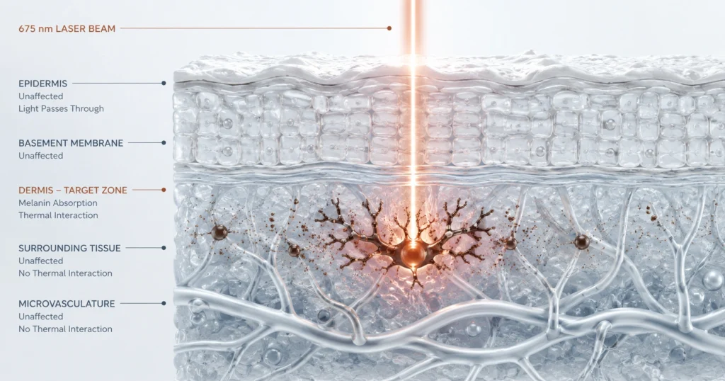
The Physiology of Structural Tightening: Understanding Thermal Denaturation and Deep Tissue Remodeling
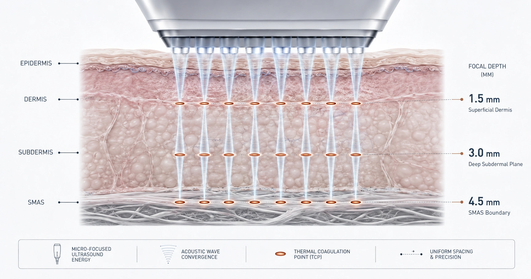
Non-surgical skin tightening using focused ultrasound technology utilizes targeted acoustic energy to induce deep tissue remodeling and manage cutaneous laxity. By establishing precise micro-injury zones at depths of 1.5 mm, 3.0 mm, and 4.5 mm, this modality initiates a controlled cellular cascade without disruptive epidermal intervention. Localized thermal elevation triggers immediate collagen fibril denaturation followed by long-term neocollagenesis. The resulting structural alignment yields documented structural adaptations that remain clinically measurable for up to one year.
The Scientific Approach to Laxity
Defining Focused Acoustic Energy in Aesthetic Medicine
In non-surgical tissue remodeling, acoustic energy functions as a highly controlled stimulus for structural modification. Global implementation of ultrasound-based tightening expanded by 13% in 2023, reflecting sustained reliance on energy-based physical modification. Micro-focused ultrasound (MFU) and high-intensity focused ultrasound (HIFU) deliver concentrated sound waves to specific subcutaneous targets while entirely bypassing epidermal disruption.
Target Layers of the Facial Architecture
Clinical protocols isolate three distinct zones for energy deposition: the deep dermis, subdermis, and the Superficial Musculoaponeurotic System (SMAS). Precise transducers utilize depth parameters of 1.5 mm, 3.0 mm, and 4.5 mm to address these structural levels directly. Focused placement ensures that structural adaptation occurs within the foundational tissue layers responsible for facial configuration.
The Thermodynamic Phase: Thermal Denaturation
Reaching the Therapeutic Temperature Window
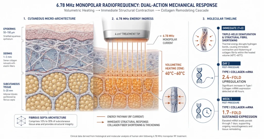
Focused ultrasound delivery elevates localized internal tissue temperatures to a precise therapeutic window between 60°C and 80°C. This thermodynamic shift relies on acoustic waves converging at a pre-calculated focal point, converting kinetic energy into thermal energy. The superficial skin layers remain unaffected by this localized internal thermal elevation.
The Biological Threshold of Collagen Fibrils
When local tissue temperatures reach the biological threshold of 57°C to 58°C, the intramolecular hydrogen bonds stabilizing collagen fibrils are disrupted. This molecular agitation results in immediate, three-dimensional collagen shrinkage and tissue contraction, a phenomenon confirmed by histological and cadaveric validation.
The Cellular Cascade of Neocollagenesis
Fibroblast Stimulation and Wound Healing Mechanics
The initial thermal phase transitions into a prolonged regenerative cycle driven by a natural wound-healing response. Transducers generate arrays of 15 to 22 discrete thermal coagulation points (TCPs) per automated line activation. These micro-injury zones stimulate prolonged fibroblast recruitment and activity within the targeted subdermal tissue.
The Timeline of Structural Synthesis
Following fibroblast activation, the biological timeline of neocollagenesis unfolds progressively over several months. Newly synthesized collagen fibers gradually mature and realign, reinforcing the structural matrix of the skin. Clinical evaluations demonstrate that these structural adaptations maintain tissue tightening and contouring benefits for up to 1 year.
Data-Driven Protocol Curation
Navigating Individual Anatomical Variance
Extensive biophysical data indicates that dermal and SMAS thicknesses vary considerably based on individual patient criteria, including age, sex, and Body Mass Index (BMI). Consequently, precise target verification is essential to ensure acoustic energy delivers to the exact intended structural depth. Curation of specific energy levels, ranging between 0.1 J and 2.5 J depending on the technical platform utilized, allows clinicians to tailor delivery parameters to match localized tissue geometry.
References
Parra AM, Dicker V, Parra LA, Martinez Amado A, Acevedo A, Castelanich D, Velasquez L. High-Intensity Focused Ultrasound Devices for Skin Tightening: A Comparative Literature Review of Device Specifications. Dermatological Reviews. 2025;6:e70030.